[ad_1]
Discussion
Scotland is in the unique position globally of having publicly funded, regular, routine eye examinations with retinal imaging for a sizeable proportion of the population (including almost everyone aged 60+) for well over a decade. In 2022/2023, for example, over 60% of 60+ year-olds in Scotland underwent a sight test in primary care optometry.25 It is important to protect such health information on behalf of the public, ensuring it is used appropriately and safely in research. Optometrists are required to keep images for 10 years; therefore, many images are already at risk of deletion, with the potential benefit to public health irretrievably lost.
Other retinal image repositories exist (eg, UK Biobank,26 Northern Ireland Cohort for the Longitudinal Study of Ageing,27 INSIGHT Health Data Research (HDR) UK,28 Age-Related Eye Disease Studies5), but SCONe benefits from Scotland’s community-acquired retinal images, including years of images prior to the emergence of signs or symptoms of disease across a broad spectrum of the population. This represents rich, longitudinal evidence not commonly available in other retinal image repositories which are mostly compiled through secondary care or disease-specific cohorts, and in some cases consist of photograph scans. There is a great deal of important information to be gleaned from the retina about changes during healthy ageing.
With the backing of the Scottish Government, we will continue to expand SCONe to capture a broad range of common and rare diseases and reflect the country’s population in terms of demographics, geography and socioeconomics. By creating a very large, representative cohort, we will maximise the potential public benefit which can be achieved through research on this invaluable resource. Crucially, participation is voluntary and at the discretion of the optometrists who are the data controllers for their patients’ images.
Strengths and limitations
SCONe is already larger than most retinal image research repositories and contains individual-level pseudonymous data from multiple linked healthcare datasets. It is stored in a secure location, with access for approved research via PHS. The current SCONe cohort reflects the sex balance in the Scottish 60+ population; it includes a slightly higher proportion of people of black and minority ethnic groups, who are disproportionately affected by eye diseases such as glaucoma and diabetic retinopathy, and a higher proportion of people from lower deprivation indices, offering the potential to explore issues of health inequality. There are hundreds of thousands of images which predate the first diagnosis of many sight-limiting and potentially life-limiting diseases.
Data routinely collected during healthcare appointments inevitably have limitations compared with a bespoke research dataset, for example, diagnostic codes (at a person, rather than an eye level) without detailed clinical findings. The purpose of data collection may be procedural rather than attempting to document a complete medical history; and with extensive longitudinal data, there are inevitably coding changes over time in response to policy updates and new targets around managing specific diseases or patient groups. Our early work is evaluating the impact of this and other issues which may lead to misinterpretation and developing guidance around taking these into account in disease prediction work. Quality assurance has been at the forefront of SCONe data collection from the outset, both in terms of interpreting existing clinical data and analysing retinal images.
Ongoing developments
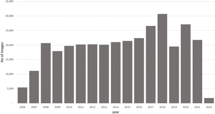
We continue to grow the repository (exploring additional dataset linkages such as the national stroke register or brain CT scans) and anticipate that we will have 1 million retinal images within the next 12 months. We will assess the feasibility of collecting optical coherence tomography or ultrawide images; however, devices which capture these are not as widespread as fundus cameras. Therefore, our main focus to achieve a large, representative cohort is on colour fundus photographs.
Inside the National Safe Haven, we are deriving metadata describing common retinal imaging phenotypes (such as vasculature, other anatomical landmarks and disease phenotypes) using automated image feature extraction methodologies and expert manual image grading to add clinically significant details unavailable from routinely collected data. We are also developing analytical methodologies to facilitate meaningful analyses which overcome the challenges inherent in real-world data described above.
Data sharing
We are conducting curation of the datasets and developing guidance to prepare them for use as a publicly accessible resource in collaboration with PHS. At the moment, access is only possible via partnership with the study sponsors. When the mechanism for broader access is established and ready to accept applications, we will publicise this. In the interim, we will publish metadata on the HDR UK gateway to allow potential users to register their interest.
[ad_2]
Source link